

A glance at a recent photo can make a Scottsdale adult notice something that wasn't obvious in the mirror, the smile looks less bright, one tooth seems darker, or yellowing appears near the gumline. That change can feel cosmetic, but tooth discoloration sometimes reflects plaque, tartar, decay, an old injury, medication exposure, or a material used during dental treatment.
Understanding what causes tooth discoloration makes the next step clearer. Some stains respond well to professional cleaning or teeth whitening. Others need bonding, veneers, a crown, periodontal care, or treatment for an underlying tooth problem. A dentist in Scottsdale can identify the pattern, examine the teeth and gums, and recommend care based on the actual cause rather than suggesting a stronger whitening product.
How a Smile Gradually Loses Its Brightness
Teeth rarely change color overnight without a reason. Coffee or tea can leave pigments on enamel, tobacco can create stubborn brown deposits, and normal aging can make the natural yellow tone of dentin more visible. A patient may also notice one darker tooth after an injury, while the rest of the smile remains unchanged.
Tooth discoloration is usually divided into surface staining and internal discoloration. That distinction helps a dental professional decide whether the next step should be cleaning, whitening, restorative treatment, or a closer evaluation with dental x-rays. It also explains why brushing harder doesn't always produce a brighter smile.
A practical starting point: The color, location, and timing of the change often provide useful clues about its cause.
The shade itself matters, but so does the pattern. Generalized yellowing may relate to surface pigments or enamel thinning. A single gray or brown tooth may require an assessment for trauma, decay, or pulpal changes. White spots, bands, or mottling can point toward developmental or mineralization changes rather than ordinary food stains.
Most discoloration is manageable once the source has been identified. The aim isn't to make teeth look whiter. A careful evaluation also checks for cavities, gum disease, worn enamel, defective restorations, cracks, and infection, so cosmetic treatment doesn't hide a condition that needs attention.
For adults searching for a dentist near me or a dentist in Scottsdale, AZ, a new-patient exam at Eric Wiitala DDS can provide that explanation in a calm, organized setting. The practice serves local patients seeking preventive, cosmetic, and restorative dental care, including people who want to understand why whitening hasn't solved a persistent shade change.
The Two Main Types of Tooth Discoloration
The simplest way to understand discoloration is to think of enamel as a painted wall. Extrinsic stains are like splatters sitting on the wall's surface. Intrinsic discoloration is more like a change in the paint itself, because the color develops within the enamel or dentin.
Dental literature has used this two-part framework for decades. Extrinsic discoloration involves pigments from food, drinks, tobacco, and other external sources collecting on the tooth surface or the acquired pellicle. Intrinsic discoloration develops from systemic, pulpal, developmental, medication-related, traumatic, or age-related changes within the tooth, as described in this clinical reference on tooth discoloration.
Why the distinction affects treatment
Surface stains often improve when a dental hygienist removes plaque, calculus, and discoloration from enamel. Teeth whitening may further lighten the natural tooth structure when the pigments are responsive to bleaching.
Internal color changes behave differently. Whitening may produce limited improvement when a stain is embedded in dentin, caused by a developmental defect, or associated with a nonvital tooth. In those cases, a dentist may discuss tooth-colored bonding, porcelain veneers, or a crown, depending on the tooth's health and structure.
A third category can blur the line. External pigments may enter through cracks, enamel defects, or worn areas and become internalized. That's why a patient can have excellent daily hygiene and still develop a localized dark area.
The following video offers a visual introduction to common discoloration patterns and causes.
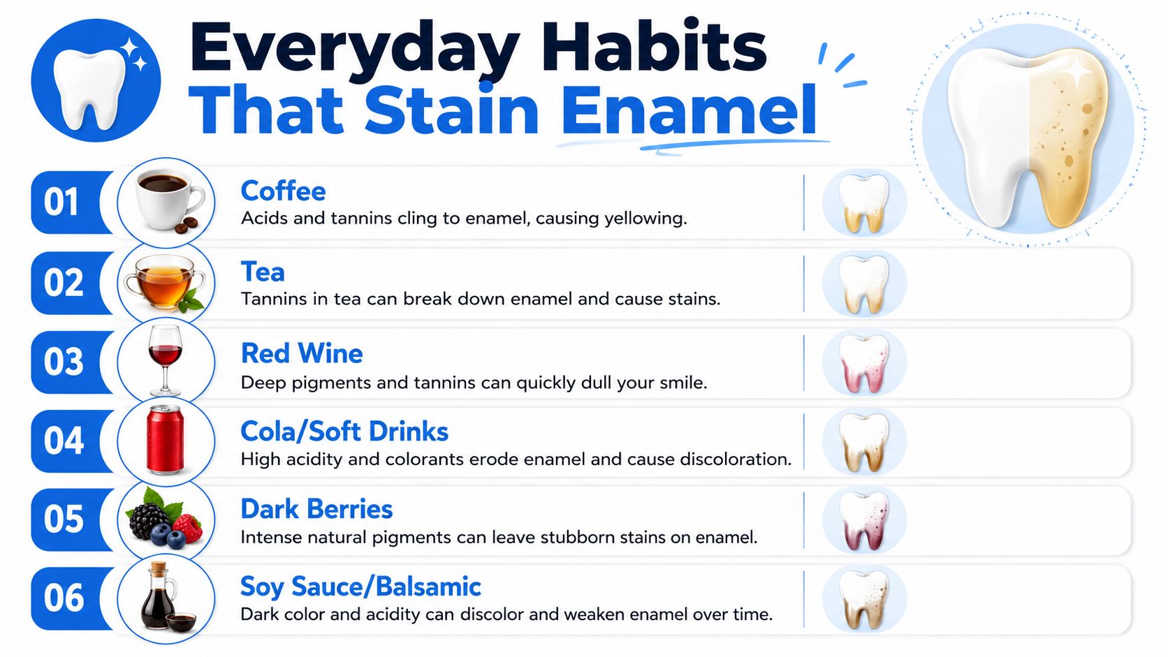
Everyday Habits That Stain Enamel
Many familiar Scottsdale routines can contribute to extrinsic staining. Coffee and tea contain chromogens, or strongly colored compounds, that can attach to the thin pellicle coating that naturally forms over enamel. Red wine, cola, dark berries, curry, and other pigmented foods can behave similarly.
The pigment usually sits more heavily in surface deposits than in the tooth tissue itself. Poor plaque control gives those compounds more material to cling to, while calculus can make discoloration at the gumline look darker and more established. The mechanism is described in this PubMed review of extrinsic tooth discoloration.
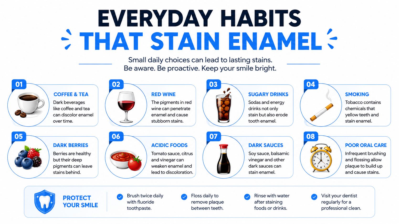
Common sources of surface pigment
- Coffee and tea: Repeated exposure allows colored compounds to collect on the pellicle, especially when plaque remains on the enamel.
- Red wine and cola: Dark beverages can leave visible deposits, and patients may notice them around crowded teeth or near the gumline.
- Berries and richly colored foods: Dark fruits, curry, and similar foods can add external pigment even when they're part of an otherwise balanced diet.
- Tobacco: Smoking and smokeless tobacco can produce persistent yellow, brown, or black deposits. Tobacco stains can be difficult to remove with home brushing alone.
- Metal salts: Iron or copper salts can contribute to extrinsic staining, whether they come from supplements or occupational exposure. The American Dental Association's whitening overview identifies metal salts as a possible source.
Some people notice staining sooner because their routine includes frequent exposure, while others accumulate pigment more slowly. Saliva flow, plaque levels, tooth position, and cleaning habits all affect how strongly chromogens remain attached.
The helpful point is that many lifestyle contributors are adjustable. A person doesn't necessarily need to eliminate every favorite drink, but changing exposure habits and maintaining professional cleanings can reduce the amount of pigment that remains on enamel.
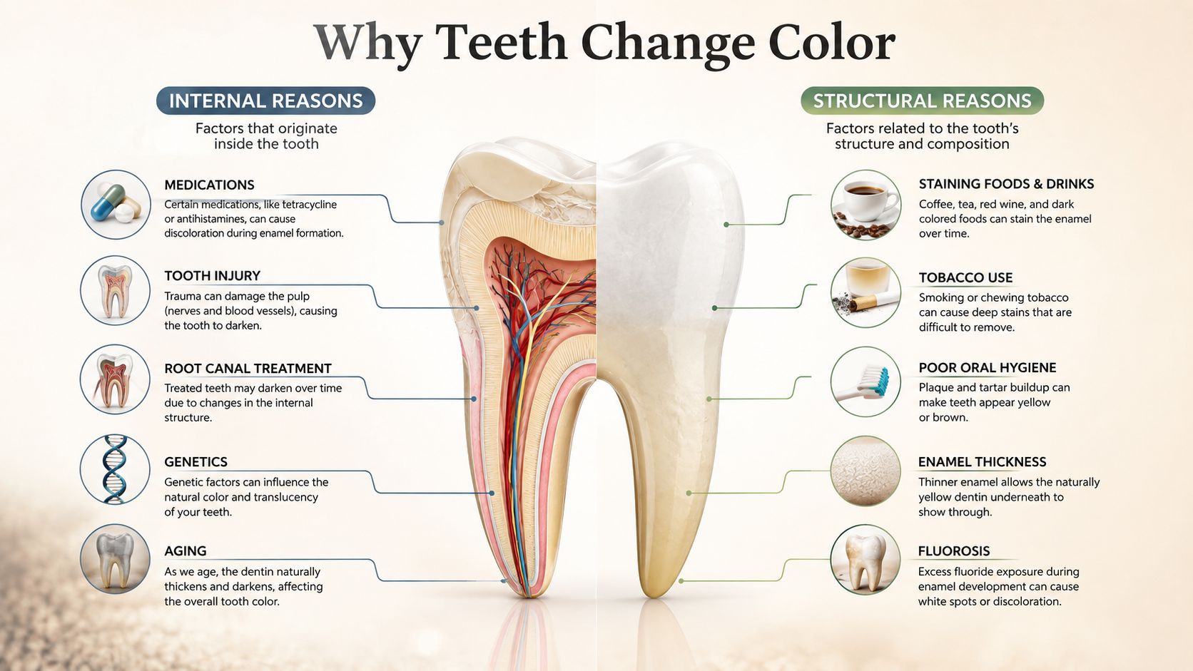
Internal and Structural Reasons Teeth Change Color
Intrinsic discoloration starts within the tooth, so a surface polish may not remove it. The shade may appear as generalized darkening, a band, a patch, or a single altered tooth rather than a uniform film across the smile.
Changes that develop over time
Aging is a common reason adult teeth look darker. Enamel gradually becomes thinner and more translucent, allowing the naturally yellower dentin underneath to show more clearly. The result can look like yellowing even when the patient's brushing routine hasn't changed.
Medication exposure during tooth formation can also alter internal color. Tetracycline exposure in childhood may become incorporated into forming mineralized tooth structure. Excess fluoride during childhood can create white opacities or brown mottling, a pattern commonly associated with fluorosis. These causes and their internal effects are discussed in this review of intrinsic tooth discoloration.
Why one tooth may look different
Trauma can damage the pulp, the living tissue inside a tooth. Bleeding or pulpal changes after an injury may later make one tooth appear gray, brown, or dark. That change deserves an examination, particularly when it follows a fall, sports injury, bite injury, or other impact.
Developmental defects can also affect color and strength from the time teeth erupt. Enamel hypoplasia and inherited conditions affecting enamel or dentin may create unusual shades, thin areas, pits, or uneven surfaces. A dentist evaluates the appearance alongside the tooth's structure, sensitivity, history, and neighboring teeth.
A useful clue: A localized band or patch often tells a different story from a generalized film across every tooth.
Intrinsic causes don't mean the smile can't be improved. They mean the treatment must match the source. Whitening may help some discoloration, while deeper changes may respond more predictably to bonding, veneers, or restorative care after the dentist confirms that the tooth is healthy.
Stains From Dental Products and Materials
A patient who brushes and flosses consistently may still see discoloration. Hygiene is important, but it isn't the only variable. Some stains result from oral-care products, dental materials, or treatment chemistry, which is why a dentist needs to ask what was used, when the color change began, and whether it affects one tooth or many.
Certain mouthwashes can leave brown or yellow surface staining with prolonged use. Chlorhexidine is a well-known example in clinical care, and some essential-oil rinses may also contribute to visible deposits. The issue isn't necessarily that the product is inappropriate. A dentist or physician may have recommended it for a specific reason, so patients shouldn't stop a prescribed rinse without professional guidance.
Root canal materials can create a different pattern. Some endodontic irrigants, sealers, temporary filling materials, and antibiotic pastes may darken a tooth from the inside out. When discoloration follows root canal treatment, a routine polishing appointment may not be enough because the material or altered tooth structure lies beneath the surface.
Why the timeline matters
A careful dental history can separate a new product stain from an older intrinsic change. Relevant details include:
- The starting point: Whether discoloration began after a new rinse, filling, root canal, medication, or injury.
- The distribution: Whether the color affects the entire smile, several teeth, or one treated tooth.
- The surface response: Whether professional cleaning changes the appearance.
- The structural clues: Whether cracks, defective restorations, or enamel defects are present.
Pigments can also enter through cracks or enamel defects, creating an internalized stain that began with an external exposure. This multifactorial pattern is recognized in the clinical discussion of tooth discoloration and treatment-related causes.
For readers comparing oral-care options, Browse Japanese toothpaste with Buy Me Japan can provide general product information, but toothpaste selection won't correct every internal or material-related stain. The right answer may be a cleaning, a change in product use, whitening, bonding, or restorative treatment.
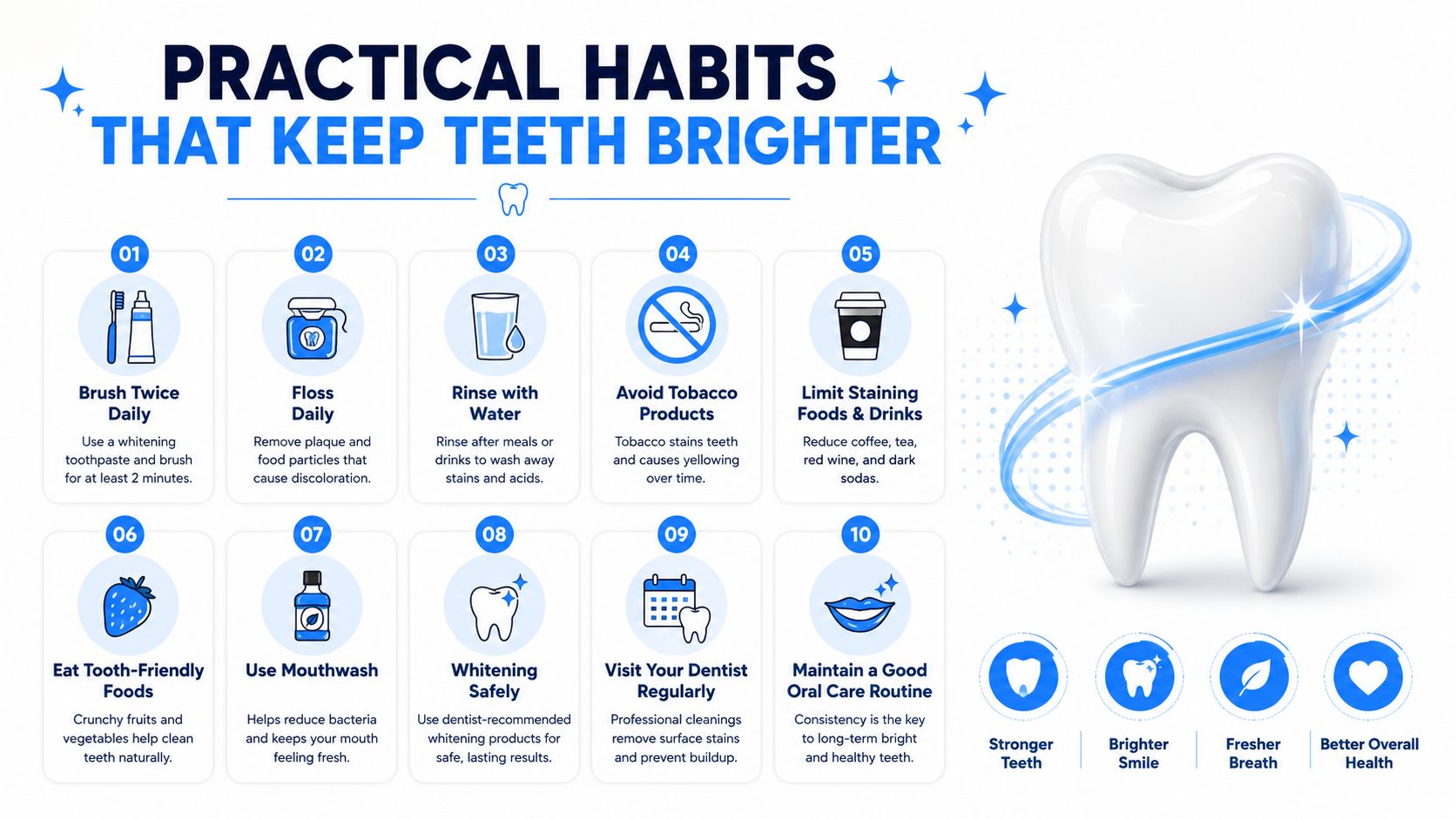
Practical Habits That Keep Teeth Brighter
Prevention works best when it fits into an existing routine. After coffee, tea, cola, or red wine, rinsing with water helps clear loose pigments from the mouth. Drinking through a straw can reduce contact between a beverage and the front teeth, while brushing or rinsing within about 30 minutes of exposure can support cleaner enamel when practical.
Daily plaque control still matters because chromogens bind more readily where plaque and calculus remain. A toothbrush, fluoride toothpaste, and cleaning between the teeth address the deposits that make surface staining look heavier. Professional hygiene visits remove buildup that home care can't reliably reach.
Small adjustments with a useful payoff
- Keep water nearby: Drinking water between meals supports a cleaner mouth and helps wash away colored residue.
- Change the exposure pattern: Sipping a dark beverage over a long period gives pigments more repeated contact with enamel.
- Address tobacco use: Stopping tobacco reduces a major source of persistent external staining and supports broader oral health.
- Schedule regular hygiene care: Consistent cleaning helps remove the pellicle-associated deposits where external pigments collect.
- Use whitening products carefully: Over-the-counter whitening products are most appropriate for responsive surface stains after a dentist has ruled out decay, gum disease, cracks, and internal causes.
Professional cleanings are commonly planned twice a year for many patients, although an individual schedule depends on oral health and periodontal risk. Patients should also use whitening products according to directions, since stronger or more frequent use isn't automatically safer or more effective.
A check-up can identify whether the discoloration is stain, calculus, enamel change, or a symptom of disease. Scottsdale patients can review preventive care and appointment expectations through Eric Wiitala DDS check-up visits.
When Discoloration Signals Something More
Not every dark spot is a cosmetic concern. A single tooth that turns gray or brown can reflect past trauma, decay, pulpal damage, or a dying nerve. Whitening that tooth before an examination may delay the care it needs and can produce an uneven result.
Discoloration near the gumline deserves attention when it appears with redness, swelling, bleeding, tenderness, bad breath, or gum recession. The dark area may be stain, but it may also be tartar or a sign of periodontal disease. A hygienist or dentist can distinguish deposits from changes in the tooth or gum tissue during an evaluation.
Patterns worth having checked
- White chalky areas: These can reflect enamel changes and may sometimes be associated with early decay.
- Brown spots: A brown area may be a cavity, especially when it catches food, feels rough, or appears alongside sensitivity.
- Dark edges around restorations: A dark patch can indicate an old restoration that has failed, leakage around the filling, or decay at the margin.
- A sudden isolated change: A recently darkened tooth should be assessed promptly, especially after an injury or dental procedure.
- Color with pain or swelling: Pain, facial swelling, drainage, fever, or difficulty opening the mouth can require urgent dental attention. Patients searching for an emergency dentist shouldn't wait for whitening when these symptoms are present.
An exam and dental x-rays at a Scottsdale office can help determine whether a shade change is purely cosmetic or connected to decay, infection, bone changes, a damaged filling, or pulp health. The evaluation also helps protect neighboring teeth, because a dark area can be easier to treat when the underlying cause is found early.
The reassuring approach is not to assume the worst and not to dismiss the change. A dentist can ask about diet, tobacco, medications, rinses, injuries, fillings, root canal care, and the timing of the color shift, then use those details to guide treatment.
Professional Treatment Options in Scottsdale
Treatment should follow the cause. A professional cleaning and hygiene visit is often the starting point for extrinsic staining because it removes plaque, calculus, and surface deposits that home brushing may leave behind. If the natural enamel remains healthy but still looks darker, teeth whitening may provide additional brightening.
Eric Wiitala DDS offers in-office whitening for patients seeking a more immediate cosmetic change, along with take-home options for gradual brightening. Patients can learn more about teeth whitening in Scottsdale during a consultation, where the dentist can assess whether whitening is appropriate for the type of discoloration present.
Matching care to the cause
- Surface deposits: Professional cleaning and improved home care may be enough when pigment sits on enamel or calculus.
- Responsive natural-tooth stains: Whitening can address certain diet-, tobacco-, and age-related color changes after an exam.
- Deep intrinsic discoloration: Porcelain veneers or dental bonding can mask color that bleaching cannot reliably lift.
- A damaged or weakened tooth: A crown may restore appearance and structure when decay, trauma, or a large defective restoration compromises the tooth.
- Missing teeth: Bridges, single-tooth dental implants, or full-arch implant solutions can restore a smile when discoloration is part of a broader restorative concern.
Bonding may also help reshape or cover a localized discolored area, while veneers can provide a more complete cosmetic change for selected healthy teeth. These options sit within broader cosmetic and restorative dentistry services that can also address chips, gaps, worn teeth, and tooth loss. Patients comparing Dental implants near me or restorative treatment should first receive an assessment of the teeth, gums, bite, and supporting bone.
A first visit generally includes a new-patient exam, a review of health and dental history, dental x-rays when indicated, and personalized treatment planning. The Scottsdale office can also discuss insurance, billing, and financing questions before treatment begins.
Eric Wiitala DDS serves patients across Scottsdale and surrounding North Valley communities. The office is located at 9755 North 90th Street, Suite B250, Scottsdale, AZ 85258, and can be reached at (480) 657-6981. Office hours are consistent Monday through Thursday, with Friday appointments available.
Eric Wiitala DDS provides professional cleanings, exams, teeth whitening, bonding, veneers, crowns, and restorative care for Scottsdale patients who want to understand and treat tooth discoloration. Schedule an appointment at Eric Wiitala DDS to identify the cause of the color change and choose a treatment plan suited to the health and appearance of the smile.